Labelled Muscles In The Body - Four Elms Primary School Monday 4th May 2020 : Ach diffuses across the gap at the neuromuscular junction and… list functions performed by skeletal mu…
Labelled Muscles In The Body - Four Elms Primary School Monday 4th May 2020 : Ach diffuses across the gap at the neuromuscular junction and… list functions performed by skeletal mu…. Bones of the foot labeling page. The musculature of 31 species was studied using phalloidin labelling and confocal laser scanning microscopy. When you are taking anatomy and physiology you will be required to identify major muscles in the human body. There are around 650 skeletal muscles within the typical human body. The movement of these muscles is directed by the learn how many muscles are in the body, how skeletal muscle attaches to bone and moves bones, and which organs include smooth muscles.
We have a lot of muscles in our bodies (literally, over 600). Blood vessels labeling page (middle school). Muscles allow us to move and function. What causes the contraction and shorten… ach released at the motor end plate. Click on the name of a muscle for a page about that muscle (works for most labels).
Muscle Map Of Human Body Muscle Map Human Body Human Anatomy Labelled Human Body Muscles Muscle Anatomy Muscle Diagram from i.pinimg.com This section explores the different types of muscles in our body and their involvement in sporting activities. View the muscles of the upper and lower extremity in the diagrams below. Muscles allow us to move and function. Labels are a means of identifying a product or container through a piece of fabric, paper, metal or plastic film onto which information about them is printed. Smooth muscle and cardiac muscle move to facilitate body functions like heartbeats and digestion. Original article on live science. The sartorius can be up to 23 inches (60 centimeters) in length, according to a 2005 paper. There are over 600 muscles in the body.
This quiz focuses on the 23 largest muscles—the ones that account for most of your mobility and strength.
Despite their similar names, teres major has different actions and innervation from the teres minor. What causes the contraction and shorten… ach released at the motor end plate. This is an online quiz called label the muscles. Ach diffuses across the gap at the neuromuscular junction and… list functions performed by skeletal mu… It stretches from the outer side of the hip bone down to the inside of the knee bone. This online quiz is called label the muscles anatomy, human, muscles, body, health, label, labeling, health science, human body. This is a table of muscles of the human anatomy. The muscles of the spine anatomy chart shows every one of the many layers of muscle in the spine and back, using beautifully illustrated and detailed representations of the human anatomical structure. This is a table of skeletal muscles of the human anatomy. We have a lot of muscles in our bodies (literally, over 600). The sartorius can be up to 23 inches (60 centimeters) in length, according to a 2005 paper. Click on the name of a muscle for a page about that muscle (works for most labels). Skeletal, or voluntary, muscles are the muscles you can control.
Muscle myofibril labeling page (advanced). Bones of the foot labeling page. Learning the major muscles of the body doesn't have to be difficult—use this anatomy quiz game to make it fun and easy! The movement of these muscles is directed by the learn how many muscles are in the body, how skeletal muscle attaches to bone and moves bones, and which organs include smooth muscles. Use the location, shape and surrounding structures to.
Muscle Labelling Help Please The Student Room from www.thestudentroom.co.uk This muscle diagram is interactive: Studying these is an ideal first step before moving labeled diagram. Two kinds of epidermal muscles are described in the palaeonemerteans and. Teres major is a thick and ovoid muscle in the upper arm. Human body muscle system, the muscles of the human body that work the skeletal system, that are under voluntary control, and that are concerned with movement, posture, and balance. Anterior muscles in the body. It stretches from the outer side of the hip bone down to the inside of the knee bone. There are over 630 muscles in the human body;
What causes the contraction and shorten… ach released at the motor end plate.
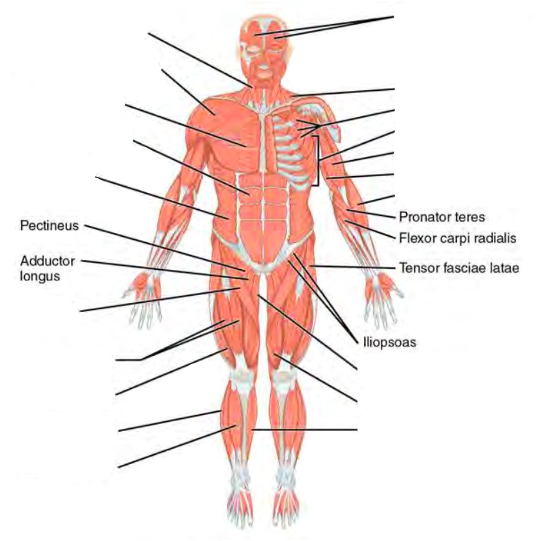
Despite their similar names, teres major has different actions and innervation from the teres minor. Smooth muscle and cardiac muscle move to facilitate body functions like heartbeats and digestion. This system is mainly concerned with producing movement through muscle contraction. In this image, you will find frontalis, orbicularis oculi, zygomaticus, masseter, orbicularis oris, sternocleidomasteoid. Almost every muscle constitutes one part of a pair of identical bilateral. Bones of the foot labeling page. This is a table of muscles of the human anatomy. Muscle anatomy quiz for anatomy and physiology! This section explores the different types of muscles in our body and their involvement in sporting activities. What causes the contraction and shorten… ach released at the motor end plate. This quiz requires labeling, so it will test your knowledge on how to identify these muscles (latissimus dorsi, trapezius, deltoid, biceps brachii. Blood vessels labeling page (middle school). Human anatomy for muscle, reproductive, and skeleton.
New data are presented on the patterns of the body wall, proboscis and gonadal musculature. **** sorry i made a mistake at 00:49 i incorrectly label and describe the thigh adductors as hip abductors. Human body muscle system, the muscles of the human body that work the skeletal system, that are under voluntary control, and that are concerned with movement, posture, and balance. Muscles, connected to bones or internal organs and blood vessels, are in charge for movement. Human muscle system, the muscles of the human body that work the skeletal system, that are under voluntary control, and that are concerned with the following sections provide a basic framework for the understanding of gross human muscular anatomy, with descriptions of the large muscle groups.
Human Anatomy Lab Manual from uta.pressbooks.pub There are approximately 640 skeletal muscles within the typical human, and almost every muscle constitutes one part of a pair of identical bilateral muscles, found on both sides, resulting in approximately 320 pairs of muscles, as presented in this article. This section explores the different types of muscles in our body and their involvement in sporting activities. When you are taking anatomy and physiology you will be required to identify major muscles in the human body. Two kinds of epidermal muscles are described in the palaeonemerteans and. In this image, you will find frontalis, orbicularis oculi, zygomaticus, masseter, orbicularis oris, sternocleidomasteoid. Types of muscles in the body. This chart includes views of the posterior thoracic wall in five separate illustrations, and almost a. Teres major is a thick and ovoid muscle in the upper arm.
What causes the contraction and shorten… ach released at the motor end plate.
Despite their similar names, teres major has different actions and innervation from the teres minor. This section explores the different types of muscles in our body and their involvement in sporting activities. It abducts and medially rotates the by and is controlled by the superior gluteal nerve. This quiz focuses on the 23 largest muscles—the ones that account for most of your mobility and strength. New data are presented on the patterns of the body wall, proboscis and gonadal musculature. Labels are a means of identifying a product or container through a piece of fabric, paper, metal or plastic film onto which information about them is printed. This quiz requires labeling, so it will test your knowledge on how to identify these muscles (latissimus dorsi, trapezius, deltoid, biceps brachii. Studying these is an ideal first step before moving labeled diagram. Original article on live science. Everyone should identify the location of skeletal muscles in the trunk and upper extremities of the. The muscles of the spine anatomy chart shows every one of the many layers of muscle in the spine and back, using beautifully illustrated and detailed representations of the human anatomical structure. Use the location, shape and surrounding structures to. This chart includes views of the posterior thoracic wall in five separate illustrations, and almost a.


Posting Komentar
0 Komentar